It is old news that damage to our brain or spinal cord is generally irreversible, leading to a gloomy prognosis for neurodegenerative disease or traumatic injury. More than a century ago, one of the founders of neurobiology, Santiago Ramon y Cajal, wrote. “In the adult centers, the nerve paths are something fixed, ended, and immutable. Everything may die, nothing may be regenerated. It is for the science of the future to change, if possible, this harsh decree.” Despite much work and substantial progress, the challenge has yet to be met.
Among many groups that have attempted to make a dent in this hard problem are those of Joshua Sanes in MCB and Center for Brain Science, and Zhigang He in the Kirby Center at Boston Children’s Hospital. In collaborative work over the past 8 years, they have used the mouse retina as a model system. Neurons in the retina called retinal ganglion cells (RGCs) send information from the retina to the rest of the brain through the optic nerve. When the optic nerve is crushed, the RGC axons are transected. Over 80% of the RGCs die within a few weeks and hardly any of the survivors regrow new axons. Why do some cells survive? Can that fraction be increased? Can rescued cells be coaxed to regenerate their axons?
The two labs bring complementary approaches to their work together. He and colleagues have discovered several interventions that improve survival and, in some cases, even promote regeneration of axons in a subset of the survivors, albeit not to a clinically useful extent. Sanes and colleagues have harnessed the power of high-throughput transcriptomic analysis (single cell RNA-seq) to classify RGCs into 46 types and identify the genes that each type expresses. In initial studies, the groups collaborated to show that types differed dramatically in their vulnerability to injury and found that manipulation of differentially expressed genes could enhance the resilience of vulnerable types (Duan et al., Neuron, 2015; Tran et al., Neuron, 2019).
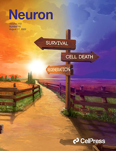
In new work, they looked more globally at gene expression patterns that affect survival and regeneration. In one of two companion papers (Jacobi et al., Neuron, 2022),(PDF), they used their transcriptomic approach to learn how three interventions discovered by the He lab act. Their analysis converged on three gene expression programs activated by injury. One appears to result in neuronal death, a second in neuronal survival and the third in regeneration of axon from surviving neurons. When axons are damaged, all three programs are activated, but the death program persists while the other two are quickly extinguished. When the retinas have been treated with ingredients from He’s recipe, the tables are turned: the death program is extinguished and the survival and regeneration programs greatly induced. The implication is that artificially enhancing expression of survival and regeneration genes could improve these qualities – an idea they validate for a few candidates.
In the companion paper, (Tian et al. Neuron, 2022) (PDF) focus on the death pathway. They perform a heroic in vivo screen of 1893 transcription factors, deleting them (using CRISPR technology) one at a time and assessing their effects on survival of injured RGCs. They found several cases in which deletion of a transcription factor enhanced survival, implying that the factor is normally part of the death program. With Dan Geschwind and colleagues at UCLA, they complemented these assays with a survey of changes in the epigenomic landscape that occur following RGC injury. These two approaches converged on a set of four key, related transcription factors, called Atf3, Atf44, C/EPBγd Ddit3, that appear to be master regulators of the choice between survival and death. Indeed, deleting these genes enhanced survival not only following injury but also in a model of glaucoma, in which blindness results from RGC dysfunction and death. Glaucoma is the most common cause of irreversible blindness worldwide and the second most common in the United States.
Together, these studies provide a global view of how neurons respond to injury, discovering novel pathways and players. Importantly, He and others have shown that mechanisms discovered in the optic optic nerve crush model illuminate processes that underlie injuries and diseases elsewhere in the central nervous system. The teams are eager to find out if this will be true for the new mechanisms and targets they have identified.
by Josh Sanes
The illustration shows the three paths an injured neuron can take – death, survival, or survival plus regeneration of a damaged axon. The horizon denotes their relative desirability. Art by Ella Marushchenko


