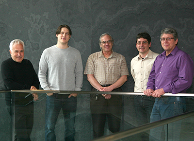
(l to r) Jeff Lichtman, Josh Morgan, Jose Conchello, Daniel Berger, and Richard Schalek
This paper describes a suite of automated technologies designed to probe the structure of neural tissue at nanometer resolution. This approach is used to generate a “saturated” reconstruction of a 1500 cubic micron sub-volume of mouse neocortex. That is, a description in which all cellular objects (axons, dendrites, and glia) and many subcellular components including 1700 synapses, more than 100,000 synaptic vesicles, dendritic spines, spine apparati, postsynaptic densities, mitochondria.
All of the data are rendered in images and itemized in a database. The approach is based on developing a means to automatically section brain tissue very thin (<30 nm), imaging it with automated scanning electron microscopy, and then aligning, segmenting and analyzing the resulting synaptic wiring.
The data set is then used to study properties of the brain’s cellular organization. In this work the synaptic connections in the cerebral cortex of a mouse are analyzed. By tracing the trajectories of all excitatory axons and noting their juxtapositions, both synaptic and non-synaptic, with every dendritic spine the data refutes the popular idea that physical proximity is sufficient to predict synaptic connectivity (the so called Peter’s rule).
Rather the analysis shows that particular excitatory axons have preferences for the spines of some dendrites more than the spines of others that they are equally in contact with. The images, segmentation, and synapse spread sheet is now an online minable database to allow general access to the intrinsic complexity of the neocortex for any researcher interested in making data-driven inquiries into the brain’s structure.
cover design by the Lichtman lab
Read more in Cell or download PDF


