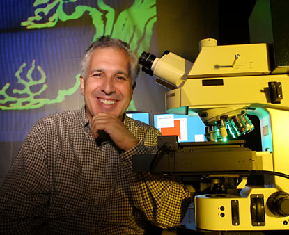
Jeff Lichtman
Double-wide color monitors glow in the darkened room like fish tanks at the New England Aquarium. Each paired screen is attached to a highly automated, computer-controlled laser scanning microscope used to watch and record the activities of fluorescently labeled nerve cells in the bodies and brains of living organisms. These remarkable machines are newcomers to MCB, housed in the Center for Brain Science’s freshly built imaging center, a cool, black-walled cavern in the Sherman Fairchild Laboratory.
The boss of the imaging center is MCB professor Jeff Lichtman, who came to Harvard last summer from Washington University Medical School in St. Louis. Lichtman has loved looking through microscopes since he was eight years old, and although he has been observing synaptic competition and elimination for two decades there is awe in his voice when he says, “We see new things, in a new way, all the time.” What sets Lichtman apart from his peers is his single-minded pursuit of descriptive neuroscience: he watches but does not interfere.
In living creatures, the brain and nervous system operate in total darkness. The availability of mice with fluorescent tags attached to various neurons and target cells, however, makes it possible to see what’s going on. These mice come from the laboratory of molecular biologist Joshua Sanes, Lichtman’s chief collaborator for the past seven years. Sanes is the director of the new Center for Brain Science, and he and Lichtman have adjacent labs. Together, they will spearhead a bold, interdisciplinary research program bent on correlating physical circuits in the brain with specific behaviors. [For more about this mission, click here.]
A typical workday for Lichtman involves managing the $2.5-million imaging center, guiding and consulting with 12 people working on a half-dozen major research projects, participating in various committees, and reviewing manuscripts for 23 different journals. Teaching will be added to his schedule next term, when he unveils a new course on optical imaging in the biological sciences. Despite all these demands on his time, lab members past and present describe Lichtman as a first-class scientist, an extremely accessible mentor, and a fine jazz pianist.
Test pilots on a tiny scale
A huge crate holding the first new microscope was waiting when Lichtman and the first wave of lab members hit campus in July. The other equipment trickled in, and today the imaging center boasts three confocal microscopes, one doubling as a multi-photon machine (with five different lasers that can excite many types of fluorescently labeled samples), and a structured illumination microscope. A dozen less exotic and less costly microscopes occupy desks and benches throughout the lab.
At Washington University, Lichtman used earlier versions of these tools to record the titanic competition of two axons, one canary yellow and the other labeled bright blue, to maintain synapses on the same target cell. It was a total surprise to find that the winning axon dances in the opponent’s end zone, forming a synapse in the very spot the loser abandoned.
More recently, postdoc Juan Carlos Tapia and his team have captured dazzling views of an axonal arbor twining around target cells like a grape vine, sprouting bunches of glowing synaptic contacts.
Lichtman Lab Members,
Past & Present
Juan Carlos Tapia
Lauren Baylor
J.D. Wylie
Bobby Kasthuri
Thomas Misgeld
Martin Kerschensteiner
Rita Balice-Gordon
It’s easy to see why Lichtman and his team are so enchanted with these top-of-the-line microscopes, even though each is as delicate and finicky as a vintage British sports car. These machines were set up by manufacturers’ representatives, each supposedly ready for action, but taken together Lichtman says they “are like the space shuttle: there are too many parts for everything to work right all at once.” Companies are eager to place their latest and greatest models in the Lichtman lab, which gives the investigators an edge in some regards but also means they are the first to stumble over bugs in machines that can cost $750,000 apiece. Nearly every day Lichtman spends time fixing something, either with his own hands or by riding herd over vendors.
One of the confocal microscopes, for example, has repeatedly gone haywire partway through a specific scanning routine that takes 30 hours. No alarms sound, so the machine finishes the run—churning out garbage while other investigators wait to use it. Eager to make things right, the manufacturer promptly put the chief software engineer and a corporate executive on an international flight to solve the problem. A recent Friday afternoon found the jet-lagged troubleshooters huddled in the eerie glow of the monitors with Tapia, the postdoc whose experiments have been hardest hit. The three talked animatedly and tinkered with a bewildering patchwork of windows crammed with grids and columns representing microscope settings.
Lichtman sat close behind the trio, pushed back in a swivel chair with his battered New Balance running shoes hooked around its base. Instead of throwing his weight around, as some lab chiefs might have done, he watched intently but let Tapia handle the situation.
Teaching birding in the dark
With people, as with neurons, Lichtman observes carefully and intervenes little. “He comes in every morning and makes rounds, almost like attending physicians do, stopping at each office along the hallway,” says Lauren Baylor, an MD-PhD student from Washington University who has been with the lab for two years. Although Lichtman averages more than one day each week on the road, lecturing and attending meetings, grad students and postdocs say they discuss their research with him nearly every day.
“If you can work independently, the lab is a wonderful environment where you can explore whatever you like. Jeff will not look over your shoulder and micromanage,” says J.D. Wylie, who recently completed his PhD. But when a lab member needs help, Wylie says that Lichtman is right there. He spends so much time meeting with lab members, in fact, that “we’ve joked about buying him one of those ‘take a number’ dispensers.”
Baylor hit a rough spot just as the lab was preparing for the move to MCB. She reached the point where she needed to know the difference between real action potentials and noise on a specific instrument, but it all looked the same to her. Lichtman spent hours teaching her how to distinguish the two. “It was like bird-watching with your advisor, learning the call of a bird and how to identify it,” she says.
Lichtman also helps students not sweat the small stuff. Former lab member Bobby Kasthuri did his masters with Lichtman before winning a Rhodes scholarship and going to Oxford for his PhD. Like many doctoral candidates, Kasthuri hit a wall and despaired of finishing his research project. He called Lichtman because “he has a very good perspective on science. He helped me see how minor things, in the grand scheme of your career, don’t matter much.” Kasthuri went on to successfully complete his PhD, and he hopes to rejoin the Lichtman team after finishing his MD in St. Louis.
Lichtman’s students also admire his speaking style. He won a dozen teaching awards at Washington University, and three current lab members sought places with Lichtman after taking his summer course on neurobiology at the Marine Biological Laboratory in Woods Hole. “We were very much impressed with how he interacted with us as students,” recalls Thomas Misgeld, an MD-PhD from Germany who has spent four postgraduate years in the lab.
Seeing is not believing
Students drawn to Lichtman’s amiable demeanor soon discover that he has a rigorous and demanding side as well. Just as a nature photographer may shoot 10 rolls of film to capture one image worth publishing in National Geographic, Lichtman estimates that he discards 100 images, sometimes 1,000, for every one he publishes or projects during a lecture. And the only way to know when you’ve got the right picture is to keep looking.
After 20 years of observing the same natural event, synapse elimination, Lichtman jokes that he is “narrow-minded as a scientist.” Students and collaborators would surely disagree: there is latitude to explore a broader range of questions, including some relevant to human diseases such as multiple sclerosis and amyotrophic lateral sclerosis.
Lichtman’s research philosophy guides all these inquiries. “We are naturalists, trying to visualize processes that have so far escaped observation,” says Misgeld. While most researchers use microscopes to “count and quantify” the results of something they’ve done, the Lichtman team uses them to explore the inner life of the nervous system much as the Hubble telescope is used to probe space. “This work requires skills that you have to learn over time. Jeff is very good at identifying details in images that you thought were odd, but you hadn’t quite picked up on them,” Misgeld says.
Lichtman’s input helped Misgeld and Martin Kerschensteiner, another MD-PhD doing postdoctoral research, discover new information about the striving of individual axons to regenerate after spinal cord injury. Kerschensteiner believes this work, which was supported by the Christopher Reeve Paralysis Foundation and has been submitted for publication, could eventually help other researchers monitor the benefits of experimental therapies for patients with spinal cord injuries.
Such results are not gained quickly or easily in the Lichtman lab, and they are never submitted for publication until they’ve been verified again and again. Rita Balice-Gordon, a postdoc from 1988 to 1994, says the most important lesson she learned from Lichtman is that “each of us should work hard to prove that our ideas are wrong—not to prove that they are right.” Now an associate professor of neuroscience at the University of Pennsylvania School of Medicine, she calls this “one of the most important lessons to learn, and one of the most difficult to teach.” It is not one that Jeff Lichtman’s students are likely to forget.


